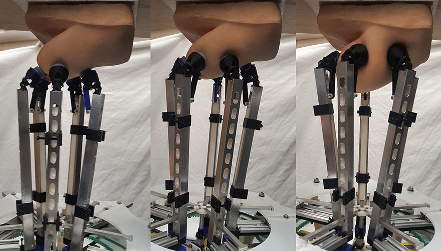
 Various stages of device examining a silicone breast constructed for the experiment. Photos: George Jenkinson
The manipulator, designed by a team at the
University of Bristol and based at the
Bristol Robotics Laboratory, is able to apply very specific forces over a range similar to forces used by human examiners and can detect lumps using
sensor technology at larger depths than ever before.
This could revolutionise how women monitor their breast health by giving them access to safe electronic CBEs (Clinical Breast Examinations), located in easily accessible places, such as pharmacies and health centres, which provide accurate results.
Precision, repeatability and accuracy are of paramount importance in these tactile medical examinations to ensure favourable patient outcomes. A range of automatic and semi-automatic devices have been proposed to aid with optimising this task, particularly for difficult to detect and hard to reach situations such as during minimally invasive surgery.
The research team included a mix of postgraduate and undergraduate researchers, supervised by Dr Antonia Tzemanaki from Bristol Robotics Laboratory. Lead author
George Jenkinson explained: “There are conflicting ideas about how useful carrying out Clinical Breast Examinations (CBEs) are for the health outcomes of the population. It is generally agreed upon that if it is well performed, then it can be a very useful and low risk diagnostic technique. There have been a few attempts in the past to use technology to improve the standard to which healthcare professionals can perform CBEs by having a robot or electronic device physically palpate breast tissue.
“But the last decade or so of technological advances in manipulation and sensor technology mean that we are now in a better position to do this. The first question that we want to answer as part of this is whether a specialised manipulator can be demonstrated to have the dexterity necessary to palpate a realistic breast size and shape.”
Simulated experimentsThe team created their manipulator using 3-D printing and other CNC techniques and employed a combination of laboratory experiments and simulated experiments on a silicone breast and its digital twin, both modelled on a volunteer at the
Simulation and Modelling in Medicine and Surgery research group at Imperial College London.
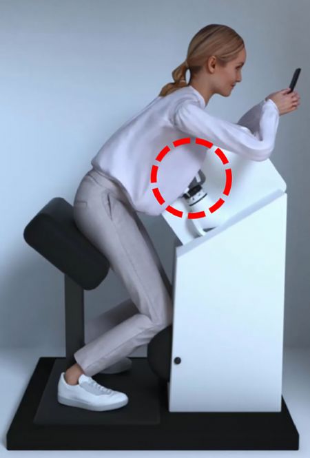 Pictured right: graphic of clinical breast examination by device
The simulations allowed the team to perform thousands of palpations and test lots of hypothetical scenarios such as calculating the difference in efficiency when using two, three, or four sensors at the same time. In the laboratory, they were able to carry out the experiments on the silicone breast to demonstrate the simulations were accurate and to experimentally discover the forces for the real equipment.
Mr Jenkinson added: “We hope that the research can contribute to and complement the arsenal of techniques used to diagnose breast cancer, and to generate a large amount of data associated with it that may be useful in trying to identify large scale trends that could help diagnose breast cancer early. One advantage that some doctors have mentioned anecdotally is that this could provide a low-risk way to objectively record health data. This could be used, for example, to compare successive examinations more easily, or as part of the information packet sent to a specialist if a patient is referred for further examination.”
As a next step, the team will combine CBE techniques learned from professionals with AI, and fully equip the manipulator with sensors to determine the effectiveness of the whole system at identifying potential cancer risks. The ultimate goal is that the device and sensors will have the capability to detect lumps more accurately and deeper than it is possible only from applying human touch. It could also be combined with other existing techniques, such as ultrasound examination.
Mr Jenkinson concluded: “So far we have laid all of the groundwork. We have shown that our robotic system has the dexterity necessary to carry out a clinical breast examination — we hope that in the future this could be a real help in diagnosing cancers early.”
This research was a part of project
ARTEMIS, funded by Cancer Research UK and supported by EPSRC.